Back to Annual Meeting Abstracts
Coil Embolization of a Ruptured Gastroduodenal Artery Pseudoaneurysm Presenting with Hemosuccus Pancreaticus.
Naveed U Saqib, Joseph DuBose, Gordon H Martin, Kristofer M Charlton-Ouw, Sheila M Coogan, Anthony L Estrera, Hazim J Safi, Ali Azizzadeh
University of Texas Medical School Houston and Memorial Hermann Heart and Vascular Institute, Houston, TX
INTRODUCTION: Hemosuccus pancreaticus (HP), hemorrhage from the papilla of Vater, is a rare form of upper gastrointestinal (GI) bleeding (incidence: 1/1,500 cases). Visceral artery pseudoaneurysms (VAPA) are secondary to chronic pancreatitis, are responsible for 20% of HP cases and can occur in up to 10% of patients with chronic pancreatitis. Gastroduodenal artery (GDA) pseudoaneurysms are among the rarest forms of VAPA (<2%). We report successful coil embolization of a ruptured GDA pseudoaneurysm in a patient with massive GI bleeding.
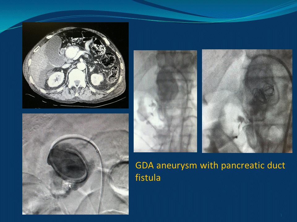
METHODS: A 68-year-old man with a history of chronic pancreatitis and cirrhosis presented to emergency room with a seven-day history of melena that was followed by acute abdominal pain and shortness of breath. On arrival, he was hypotensive with severe anemia (Hemoglobin 4.2). The patient was aggressively resuscitated with transfusions. Next, an esophagoduodenoscopy was performed, which did not reveal the source of bleeding. A computed tomographic angiography (CTA) of the abdomen and pelvis demonstrated a 4 cm GDA pseudoaneurysm without active extravasation. The patient was taken to the operating room for an aortogram and possible coil embolization.
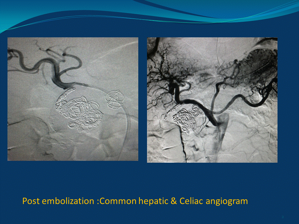
RESULTS: A transfemoral selective common hepatic artery and superselective gastroduodenal angiogram was performed. The angiogram revealed a large GDA pseudoaneurysm with a pancreatic duct fistula as the source of the GI bleed. Contrast extravasation into the duodenum was demonstrated on delayed images. A 6 Fr sheath (Pinnacle® Destination, Terumo) was placed in the main trunk of celiac artery, a 5 Fr catheter (Glidecath®, Terumo) was placed in the common hepatic artery, and a 3 Fr microcatheter (Renegade®, Boston Scientific) was placed in the aneurysm sac. The aneurysm sac was packed with Interlock™ Fibered IDC™ (Boston Scientific) coils delivered via the microcatheter. Subsequently, the GDA was embolized with Interlock™ Fibered IDC™ (Boston Scientific) 3 mm coils. Post-embolization common hepatic angiogram revealed successful exclusion of the sac, flush occlusion of the GDA, and patent hepatic arteries. Selective celiac and SMA angiogram revealed patent vessels with no retrograde flow into the VAPA. The patient had an unremarkable postoperative course without any further bleeding. He was discharged on postoperative day 5. A follow-up CTA confirmed continued exclusion of VAPA.
CONCLUSION: VAPA should be included in the differential diagnosis of patients presenting with GI bleeding. Coil embolization using micro-catheter techniques is a suitable treatment option for this challenging clinical condition. Advanced embolization techniques should be included in the training and practice of modern vascular surgeons. 

Back to Annual Meeting Abstracts
