
Primary Tear Distance From Left Subclavian Artery Predicts Growth in Chronic Type B Aortic Dissection
Jesse A Codner, Xiaoying Lou, Yazan M Duwayri, William D Jordan, Jr., Edward P Chen, Bradley G Leshnower
Emory University School of Medicine, Atlanta, GA
Background. The optimal treatment for acute uncomplicated type B aortic dissections (auTBAD) remains controversial. Currently, thoracic endovascular aortic repair (TEVAR) is indicated for the onset of complications. This study investigates whether radiographic characteristics of the primary intimal tear (PIT) in auTBAD can predict the onset of delayed aneurysm formation.
Methods. A review of a U.S institutional aortic database from 2000 to 2016 identified 318 auTBADs initially treated with optimal medical therapy. From this cohort, 103 patients with two CT or MRI scans > 6 months apart were available for imaging analysis and were included in this study. These patients were divided into subgroups based upon growth of the thoracic aorta ≥ 1 cm: (1) No Growth (n=46) and (2) Growth (n=57). Twenty-five patients (43.9%) in the Growth group underwent open or endovascular intervention. TeraRecon (Foster City < CA) imaging software was used to analyze characteristics of the PIT including the maximum width, maximum length, and distance from the left subclavian artery (LSA) of the primary tear. Statistical comparisons between groups were performed using Chi-Square, Fisher, Mann-Whitney and t-tests.
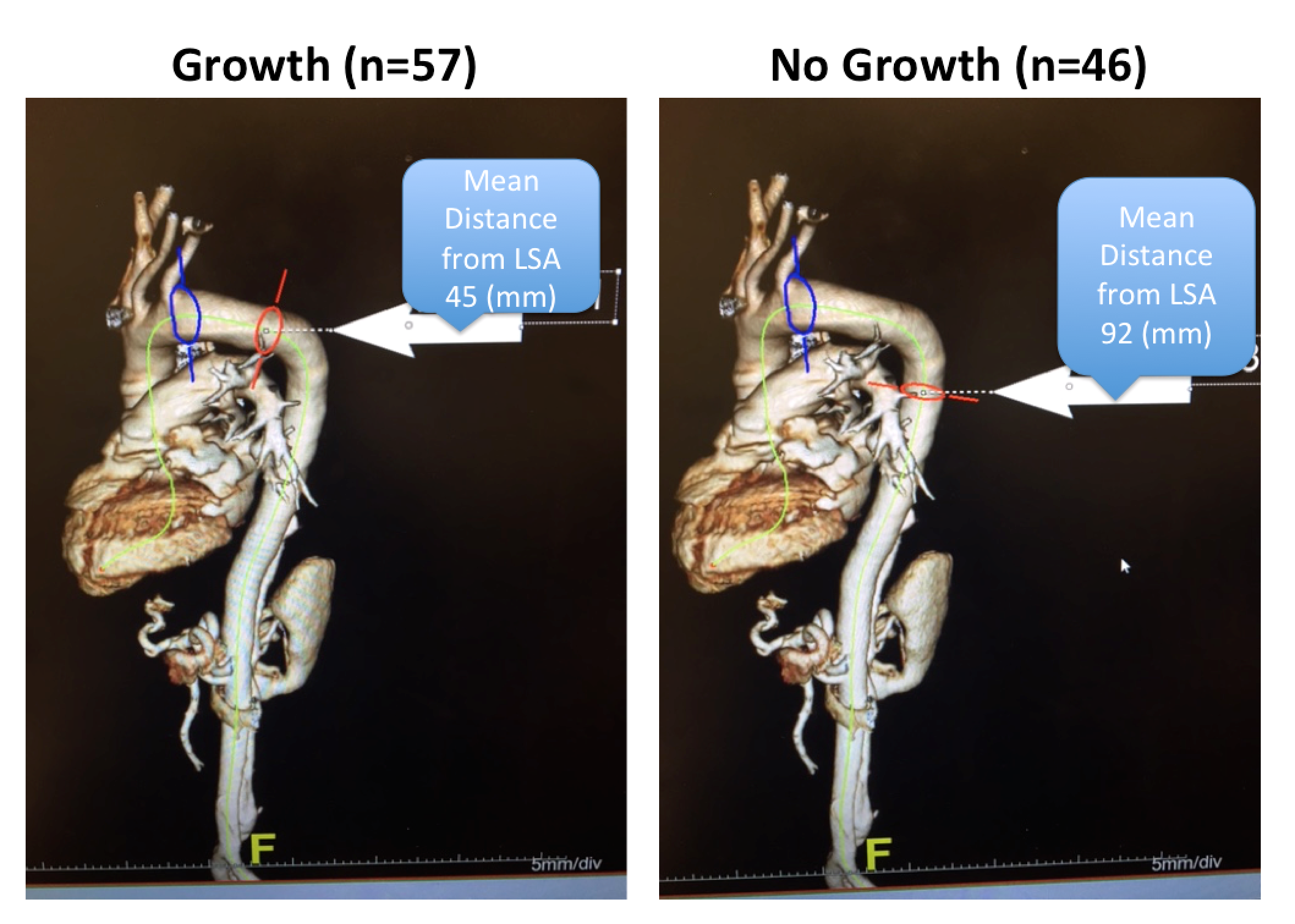
Results. The mean age of all patients was 53 ± 11 and 70% were male. There were no differences between groups in age, gender, hypertension, diabetes mellitus, tobacco abuse, COPD or renal failure. The mean follow-up was equivalent between the two groups (Growth 33 ± 27 months vs No Growth 40 ± 34 months, p=0.3). There was no difference in the maximum diameters of the thoracic (Growth 4.4 ± 0.9 cm vs No Growth 4.2 ± 0.7 cm, p=0.16) or abdominal (Growth 3.7 ± 1.1 cm vs No Growth 3.5 ± 0.5 cm, p=0.38) aorta at the time of presentation between the two groups. The distance of the primary intimal tear from the left subclavian artery in patients with auTBAD was significantly shorter in the Growth group compared to the No Growth group (Growth 45 ± 48 mm vs No Growth 92 ± 78 mm, p=0.001, Figure). There was no difference in the maximum length or width of the primary intimal tear between the two groups. (Table).
Conclusion. The distance of the primary intimal tear from the left subclavian artery predicts the development of descending thoracic or thoracoabdominal aneurysms in the chronic phase of TBAD. Patients with primary tears located in the distal arch (Zone 3) should be monitored more closely and may be considered for early TEVAR in the setting of acute uncomplicated Type B aortic dissection.
| Radiographic Characteristics | Growth (n=57) | No Growth (n=46) | P-Value |
| PIT Distance from Left Subclavian Artery (mm) | 45.4 +/- 48.1 | 91.5 +/- 77.8 | 0.001* |
| PIT Maximum Width (mm) | 11.3 +/- 6.6 | 10.0 +/- 6.9 | 0.33 |
| PIT Maximum Length (mm) | 11.7 +/- 11.0 | 9.5 +/- 8.9 | 0.28 |
| Max Diameter of Thoracic Aorta at Presentation (cm) | 4.4 +/- 0.9 | 4.2 +/- 0.7 | 0.16 |
| Max Diameter of Abd Aorta at Presentation (cm) | 3.7 +/- 1.1 | 3.5 +/- 0.5 | 0.38 |
Back to 2018 Abstracts


