
The Epidermal Growth Factor Pathway Axis is Disrupted in Diabetic Mesenchymal Stem Cells from Peripheral Arterial Disease Patients
Tatiana Chadid1, Andrew Morris1, Alexandra Surowiec1, Scott Robinson2, Katie Kuo3, Haiyan Li3, Mason Griffin4, Jacques Galipeau5, Luke P Brewster1
1Department of Surgery, Emory University School of Medicine, Atlanta, GA;2Department of Surgery, University of Michigan, Ann Arbor, MI;3Department of Surgery, Emory University School of Medicine,, Atlanta, GA;4University of Georgia, Atlanta, GA;5University of Wisconsin in Madison, Madison, WI
Introduction: Diabetes impairs vascular regeneration accelerating diseases of aging including peripheral arterial disease (PAD). Diabetic patients develop PAD ~15 years earlier than non-diabetic patients, and they have a much higher risk of major amputation than the non-diabetic population (>8x). PAD patients have poor blood flow to their leg muscles resulting in muscle loss, leg pain (ischemic myopathy), and in diabetic patients, this often leads to major amputation. Pathologically, there are profound defects in the vascular supply of PAD muscle with subsequent damage to the skeletal muscle. Such defects overwhelm ones’ innate regenerative capabilities. While the number of therapeutic options for PAD have improved, diabetic patients continue to require a disproportionate number of major amputation. Thus diabetic patients with PAD are in urgent need of regenerative therapies. Mesenchymal stem cells (MSC) can be used from the bone marrow (BM-dMSCs) or adipose tissue of patients (ATD-dMSCs), and they are powerful tools for regenerative medicine. While MSCs may differentiate into many cell types in vitro, in vivo human MSCs have limited differentiation capacity and function as a pericyte-like cell promoting regeneration through stromal and paracrine pathways in targeted tissue beds. Unfortunately in clinical trials for PAD, diabetic patients do not appear to benefit from cell therapies like their non-diabetic counterparts do. While this may be due to intrinsic defects in diabetic mesenchymal stem cells’ (dMSCs) regenerative capacity (reported in rodent models), recent patient data from our laboratory and others suggest this is not so. Still diabetic MSCs do have limitations in proliferative capacity that may affect cell survival (and ultimate utility for PAD patients). Since epidermal growth factor (EGF) is known to be important to cell proliferation and survival pathways, we hypothesized that dMSCs have EGF signaling defects that may be repaired by targeted culture supplements. The objective of this work was to determine the mechanism of dMSC dysfunction in human dMSCs and the regenerative capacity of dMSCs from both bone marrow and adipose-tissue reservoirs.
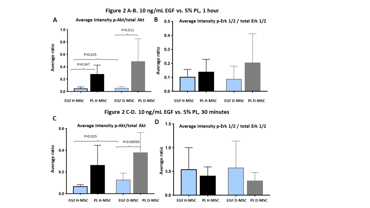
Methods: dMSCs were cultured from the bone marrow and were initially stimulated for 24 hours with either standard MSC culture media, 5% fetal bovine serum + 25 ug/mL fibroblast growth factor-2, or 5% human platelet lysate (PL), which is replete with EGF. We then compared EGF (25 ug/mL) supplementation of 5% FBS to 5% PL at 30 minute and 1 hour time points. Western blot analyses were then performed on dMSC cell lysates for pEGFR and EGFR; pERK1/2 and ERK 1/2; and pAKT and AKT. GAPDH was the control protein. Healthy donor MSCs (H-MSC) were used as control cells.
The regenerative capacity of dMSCs from bone marrow and adipose tissue from 5 patients (6 limbs) was determined by secretome analyses and in vitro stimulation of human umbilical vein endothelial cells (ECs). The secretion of angiogens by dMSCs including hepatocyte growth factor (HGF), vascular endothelial growth factor (VEGF), monocyte chemoattractant protein-1 (MCP-1) and epidermal growth factor (EGF) was quantified using Luminex technology. The mean fluorescence intensity (MFI) of each protein was averaged for all patients and the relative increase of each protein in the conditioned media was calculated. An increase of 1.4 over background was considered significant and culture media without cells served as the background control. dMSC’s mitogenic activity was tested by culturing ECs in media from either BM- or ATD-dMSCs and proliferation was captured fluorescently. dMSCs’ chemotactic activity was quantified using a modified Boyden chamber by platting the ECs with pooled conditioned media (CM) from either BM-dMSCs or ATD-dMSCs and the number of EC that migrated were counted in a blinded fashion. Results were compared to positive control (EC growth media) and negative control (quiescent media). A 3-D co-culture EC sprouting assay was performed by co-culturing PKH-stained ECs with bone marrow-MSCs or Adipose tissue derived- MSCs and trapping the cell pellets in a fibrin hydrogel. EC sprouting was quantified by fluorescent microscopy and expressed as a ratio of EC sprouting in co-culture with MSCs divided by that of the EC-alone pellet. To test dMSC angiogenic activity under diabetic conditions, dMSC:EC co-culture assays were performed under hyperosmolar conditions [25mM glucose or mannose]. Paired t-test, ANOVA and Kruskal-Wallis tests were used as appropriate.
Results: Under standard culture conditions, dMSCs had diminished pAKT response to FGF-2 supplemented media compared to H-MSCs. PL supplementation recovered pAKT levels in dMSCs (Figure 1A). In contrast, pERK 1/2 levels were stimulated by PL in H-MSCs but not dMSCs (Figure 1B). Next, we determined the EGF dose-response curve in H-MSCs. Here we found increasing pEGFR/EGFR ratios with increasing dosages (Figure 1C), but that 10 ng/mL EGF had the optimal pERK1/2:ERK1/2 ratio in H-MSCs (Figure 1D). In order to determine whether EGF supplementation was sufficient for recovery of AKT and ERK 1/2 phosphorylation in dMSCs, we compared 10 ng/mL of EGF to 5% PL in both dMSCs and H-MSCs. Here we found that pAKT:AKT ratios were significantly increased by PL (over EGF alone) in both H-MSC and d-MSCs at 30 minute and 1 hour time points (Figure 2A/C), but that pERK 1/2:ERK 1/2 ratios were similar between groups at these time points (Figure 2B/D.
The dMSC secretome had robust levels of the angiogens that were similar for both bone marrow and adipose tissue-derived dMSCs: VEGF (fold increase of 22 ±8.9 and 21.6 ± 9.3), MCP-1 (fold increase of 69.2 ± 40 and 62.4 ± 36.3), and HGF (Fold increase of 7.6 ± 6.7 and 5.7 ±3.6). Interestingly, EGF secretion by both bone marrow and adipose tissue-derived dMSCs was negligible in the secretome (0.89 and 0.91). Similarly, the conditioned media (CM) from both bone marrow (BM) and adipose tissue-derived (ATD) dMSCs stimulated similar HUVEC proliferation that was significantly greater than quiescent media (QM) [1.08 nm ± 0.29 nm for BM-MSC (P=.01) and 0.95 nm ± 0.21 nm for ATD-MSC (P=.04) vs. 0.41 nm ± 0.05 nm for QM] and similar to that of EC growth media (BM-MSCs vs. EC media, P=0.9; ATD-MSCs vs EC media, P=0.8). Similarly, CM from BM- and ATD-dMSCs stimulated significantly greater EC migration over that of QM [(196.8 cells ± 32.6 cells for BM-MSCs; P=.007) and (208 cells ± 36.3 cells for ATD-MSCs; P=.005) vs. 114.8 cells ± 53.3 cells for QM; This too was similar to the migration stimulated by EC growth media (P=.9 and P=.8, respectively). In order to test dMSC angiogenic effect on ECs, a co-culture assay (1:1 dMSC:EC) was performed. Here, co-culture with ECs led to significantly longer EC sprouts than EC-alone cell pellets [Day 3: (148.4 µm ± 69.8 µm for BM-MSC/EC; P<.05) and (157.8 µm ± 76.6 µm for ATD-MSC/EC; P<.05); EC only pellet: 52.8 µm ± 49.7 µm. Only under diabetic conditions were there significant differences between BM and ATD-dMSCs. Here, ATD-dMSCs promoted greater EC sprouting lengths than BM-dMSCs at day 3 under both the 25mM glucose and mannose conditions (P=.0029; P=.0001, respectively).
Conclusions: The EGFR axis in dMSCs from PAD patients appears to be defective. This is likely due to a deficiency in EGF secretion by dMSCs that requires exogenous supplementation. Our results support that EGF supplementation may help overcome dMSC proliferation and survival cascades, but that AKT pathways may be further benefitted by PL over EGF alone. Since our data supports robust angiogenic activity of both bone marrow and adipose tissue-derived dMSCs, exogenous EGF or PL strategies (including gel impregnation) may be an effective strategy for not only improving dMSC expansion but also prolonging dMSC survival for cellular therapies. Finally, if our observed benefit of ATD-dMSC over BM-dMSCs under diabetic conditions is subsequently validated, the choice of MSC reservoir may also be of use in optimizing cell therapies for diabetic patients with PAD. 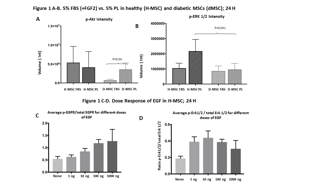
Back to 2018 Abstracts


