
The Effects of Glucose Starvation and B27 Depletion on the Expression of Erythropoietin Receptor in Neurons
Mohamed Eldeiry, Katsuhiro Yamanaka, Muhammad Aftab, Joseph C. Cleveland, Jr., Thomas Ryan, Xianzhong Meng, Michael J. Weyant, David A Fullerton, T. Brett Reece
University of Colorado Denver, Aurora, CO
INTRODUCTION: Spinal cord ischemia is a dreaded complication of complex aortic repairs that can result in paraplegia in up to 10-20% of cases. Erythropoietin plays an important, but still unclear, role in protection of the spinal cord during ischemic injury. This role is elucidated by heterodimerization of the erythropoietin receptor (EpoR) with the beta common receptor (βCR) as previously demonstrated in a murine model of spinal cord ischemia-reperfusion injury. This was observed along with increased activation of signal transducer and activator of transcription 3 (STAT3). However, the cell-specific role and mechanism of βCR and EpoR upregulation and signaling remain to be elucidated. The current model of neuronal injury employs oxygen and glucose starvation along with B27 neuronal supplement deprivation from the cell culture media. We hypothesize that in vitro glucose starvation will lead to upregulation of BCR expression and wanted to further evaluate the role of B27 depletion.
METHODS: Neurons were obtained from 2-4 day old neonatal mice. Cortical tissue was separated and enzymatically digested and cells were then separated using a density gradient. Neuronal fraction was obtained and the cells were seeded onto polymer coated 48-well culture plates. The cells were cultured in serum free media supplemented by B27 as previously reported. After reaching maturity in 5-7 days the neurons were treated with media that was either glucose or B27 depleted for 1, 2, and 4 hours. Cells were lysed and Western Blot analysis was used to evaluate the βCR and EpoR expression along with STAT3 activation in the lysate.
RESULTS: Glucose starvation resulted in a time-dependent increase in βCR expression peaking at 2 hours (3.0 ± 0.7 vs. 1.0 ± 0.4, p < 0.05) (Figure a). Alternatively, B27 deprivation did not affect βCR expression (Figure a). Neurons deprived from B27 had no detectable expression of EpoR (Figure b). The activation of STAT3 peaked at 1 hour in both glucose starved and B27 deprived neurons (3.8 ± 0.6 and 3.7 ± 0.6 vs. 1.0 ± 0.6, p < 0.05). Subsequent activation of STAT3 decreased over time but seemed to persist above baseline (Figure c).
CONCLUSIONS: We evaluated the role of glucose starvation along with B27 supplement deprivation in neurons as a surrogate of spinal cord ischemia reperfusion. Glucose starvation causes a time-dependent increase in βCR expression without affecting EpoR expression. On the other hand, B27 deprivation doesn’t affect βCR expression but does lead to undetectable EpoR expression, possibly related to receptor shedding. STAT3 activation peaked early in the experiment and preceded maximal βCR expression, suggesting that while STAT3 activation may play a role in neuronal injury, it may work parallel to βCR and its mechanisms of protection. These data give insight into the model of neuronal injury which can be used to further guide study of this complex neuronal network as it relates to spinal cord protection. 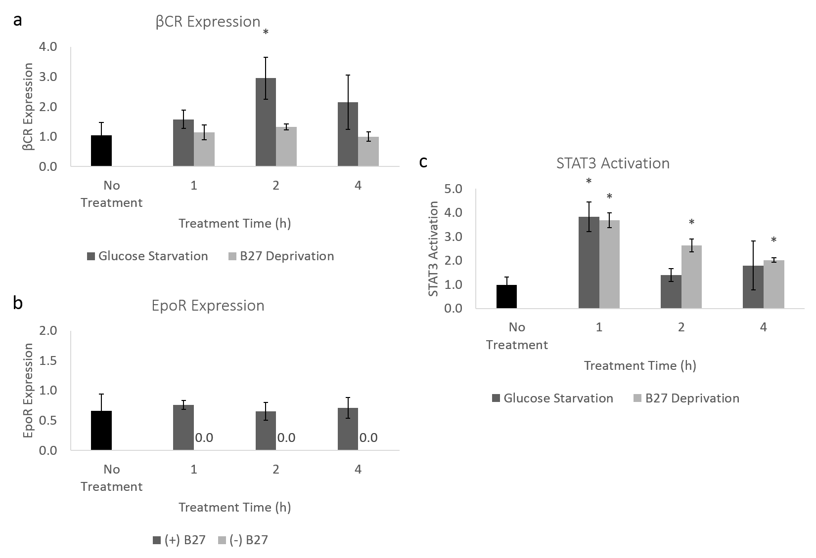
Back to 2018 Posters


