
Dual Fluoroscopy with Live-Image Zooming Significantly Reduces Patient and Operating Staff Radiation During Fenestrated/Branched Endovascular Aortic Aneurysm Repair (F-BEVAR)
Laura Timaran1, Carlos Timaran2, Carla Scott2, Marilisa Soto-Gonzalez2, David Timaran-Montenegro2, Melissa Kirkwood2
1University of Pittsburgh, Pittsburgh, PA;2UT Southwestern Med Ctr, Dallas, TX
Background: Fenestrated/branched endovascular aneurysm repair (F-BEVAR) is a complex procedure that generates high radiation dose. Utilizing magnification views aids in the success of vessel cannulation but increases the overall radiation dose. The aim of the study was to compare the radiation doses to patients and operating room staff from two fluoroscopic techniques, standard magnification vs. dual fluoroscopy with live-image digital zooming during F-BEVAR.
Methods: An observational, prospective, single center study of F-BEVAR procedures using Philips Allura XperFD20 equipment was performed over a 42- month period. Intravascular ultrasound, 3D-fusion and extreme collimation were used in all procedures. Intraoperative live imaging processing was performed using two imaging systems: standard magnification in 123 (81%) and dual fluoroscopy with live-image digital zooming in 28 patients (18%). In the latter, the live ‘processed’ (zoomed/subtracted) images are displayed on examination monitors and live images are displayed on reference monitors. The reference air kerma was collected and represents patient dose. Operating staff personal dosimetry was collected using the DoseAware system (Philips Healthcare, Amsterdam, The Netherlands). Patient and staff radiation doses were compared using non-parametric tests.
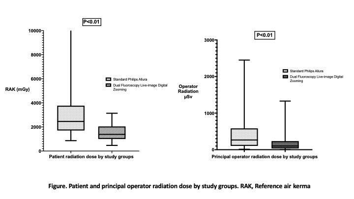
Results: Mean age was 71.6 ± 11.4 years. The median BMI was 27 kg/m2 (Interquartile range [IQR], 24.41- 30.55) and was the same for both groups. Procedures performed with dual fluoroscopy with live-image zooming demonstrated significantly lower median patient (1382 mGy [IQR, 999-2045] vs 2458 mGy [IQR, 1706-3767 mGy]) (P<0.01) and primary operator radiation doses (101 μSv [IQR, 34-235 µSv] vs 266 μSv [IQR, 104-583 µSv]) (P<0.01) when compared to standard magnification (Fig). Similar significantly reduced radiation doses were recorded for first assistant, scrub nurse and anesthesia staff in procedures performed with dual fluoroscopy (Table). According to device design, procedures performed with 4-fenestration/branch devices generated higher operator radiation doses (262 µSv [IQR, 116.5-572 µSv] vs 171[IQR, 44-325 µSv]) (P<0.01) compared to procedures with 3 or less fenestration/branches. Among the most complex design (4-vessel), operator radiation dose was significantly lower with dual fluoroscopy compared to standard imaging (59.5 µSv [IQR, 19.5-155 µSv] vs 309 µSv[IQR, 150-611 µSv]) (P=0.01)..
Conclusions: Current radiation doses to patients and operating personnel during F-BEVAR are within acceptable limits. Dual fluoroscopy with live-image zooming, however, results in dramatically lower radiation dosages compared to the standard image processing and magnification. Operator radiation doses were up to 5 times lower during procedures performed using more complex device designs when dual fluoroscopy was used.

Back to 2020 Abstracts