
Management of a Large Symptomatic Portal Vein Aneurysm
Jeffrey B Edwards1, Lila B Cohen1, Amy Lu2
1University of South Florida, Tampa, FL;2Tampa General Hospital, Tampa, FL
INTRODUCTION: Visceral venous aneurysms are rare. Though often related to portal venous hypertension, they can also occur in otherwise healthy individuals and may present with abdominal pain, rupture, or thrombosis.
METHODS: An otherwise healthy female presented with nine-months of abdominal pain. Workup for gastrointestinal and hepatobiliary conditions was unremarkable, and computed tomography (CT) scan revealed 4-cm aneurysm at the portal venous confluence. Here we describe her surgical management.
RESULTS: A 42-year-old female smoker presented with nine months of abdominal pain with associated nausea, vomiting, and diarrhea. These symptoms had been gradually increasing in severity, resulting in a 35 pound weight loss. Pertinent medical history included compensated congestive heart failure, asthma, and thyroid cancer. She had previously undergone a thyroidectomy, hysterectomy, and cholecystectomy. She occasionally used marijuana and had a remote history of amphetamine use. Workup include esophagogastroduodenoscopy and colonoscopy which revealed gastritis and duodenitis with a benign 4mm polyp in the ascending colon. Computed tomography (CT) scan of the abdomen revealed a 4cm aneurysm at the portal venous confluence (Image 1). Liver Doppler confirmed patency of the portal vein and did not reveal any features of cirrhosis. There was a normal portosystemic pressure gradient as measured by transjugular hepatic venogram. Due to her abdominal symptoms and concern for rupture, the patient elected to proceed with surgery. Through a midline laparotomy, the porta hepatis was controlled and the aneurysm was exposed. We obtained inframesocolic control of the SMA and with top-down and medial-lateral dissection of the aneurysm we obtained proximal control of the splenic, portal, and superior mesenteric veins. We dissected the aneurysm free of the pancreas, which was displaced antero-inferiorly. Tangential aneurysmectomy and aneurysmorrhaphy was performed to restore the normal portal venous confluence (Image 2). She was discharged on post-operative day 4 with no complications and has remained pain free and asymptomatic. Post-operative CT scan revealed good result with patent repair.
CONCLUSIONS:Visceral venous aneurysms are rare and available data is limited to case reports and small series. While therapy must be tailored to each individual patient, diagnosis can be easily made with contrasted CT imaging and surgical resection with primary reconstruction is a safe, viable option. 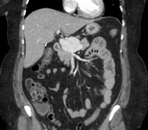

Back to 2020 Abstracts