


Computer vision analysis of clinic video for enhanced risk and resilience screening.
Chloe A Powell1, Meghan R Dailey1, Melina Varlamos1, Christopher Brown1, Akul Arora1, Vanessa Niba1, Neal Duggal1, Michael R Mathis1, Matthew P Goldman2, Matthew A Corriere1
1University of Michigan Health System, Ann Arbor, MI;2Wake Forest School of Medicine, Winston Salem, NC
INTRODUCTION: Consensus guidelines exist for perioperative risk assessment before major vascular procedures, but risk assessment often remains a subjective and inconsistent process. Frailty and comorbidity are well-characterized predictors of adverse perioperative outcomes, yet most evidence related to these risk factors is retrospective. We developed an artificial intelligence approach to risk screening based on computer vision analysis of video data to permit automated risk screening.
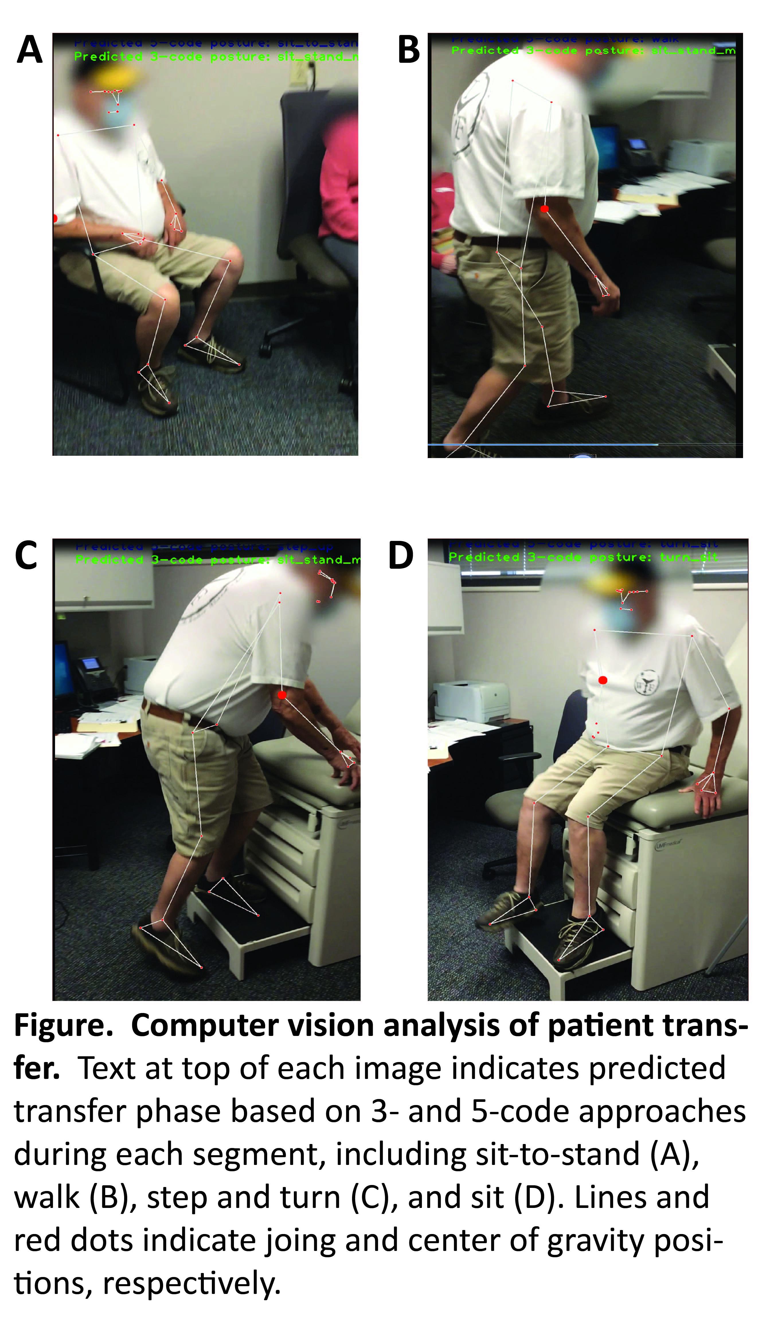
METHODS: Patients evaluated in vascular surgery clinics were prospectively recruited. Data collected included: 1) a video of the patient moving from a chair to the examination table; 2) demographic and comorbidity data; and 3) dominant hand grip strength. Video data were manually coded and segmented into transfer phases based on the Timed Up and Go (TUG) Test, a validated measure of fall risk and mobility. Video time stamps were used to quantify transfer phase durations.. Videos were uploaded into a machine learning pipeline, split into frames to process individually, and passed into the MediaPipe BlazePose Machine Learning framework to output a “landmark” pose, which includes the xy location of 33 human features and joints. We also predicted the center of gravity from these output locations. Locations and predicted center of gravity were projected onto the video to track patient movement. Movement data were correlated with labeled temporal data to train a machine learning model to classify segmented tranfer phases based on the timed up-and-go test using random forest and support vector machine (SVM) approaches.
RESULTS: 100 patients were recruited. Random forest classification was superior to SVM modeling, and was 98% accurate for classification of segmented transfer phases (Figure). Factors associated with systematic errors included multiple people in the frame (e.g., a family member in addition to the patient) and inanimate objects in chairs (e.g., handbags) prior to initiation of the transfer task. Classification accuracy was not reduced by patient face blurring for deidentification, landscape versus portrait orientation during video recording, or room-specific factors (including room layout, type of exam table, and chair orientation).
CONCLUSIONS: Automated analysis of patient movement and appearance has potential to improve preoperative risk evaluation. Use of video data for risk screening also has potential to overcome limitations related to missing data at the time of treatment selection, including screening through uploaded videos submitted by patients before in-person appointments. Next steps will be correlation of movement data with clinical data, and qualitative factors related to patient appearance, and outcomes.
Back to 2024 Abstracts